The Diaphragm and Hiatal Hernias
by Robert Tallitsch, PhD | April 14, 2025

Case study video explaining the diaphragm and hiatal hernias with a patient example!
Use the button below to schedule a demo to learn about our quizzes, flash cards, and other anatomy resources that support this Brain Builder!
Schedule a Demo
A hernia is defined as the protrusion of fatty tissue, an organ, or a part of an organ into or through a weak spot in either the muscular tissue or connective tissue that composes part of the abdominal wall or diaphragm. Some of the most common types of hernias are:
- Epigastric hernia: This type of hernia occurs when fat or other abdominal contents push through a weakened segment of the wall of the abdomen in the epigastric region of the abdomen. (The epigastric region is the upper, central part of the abdomen between the umbilicus and rib cage.)
-
Hiatal hernia: Here, a segment of the esophagus and or stomach pushes through the hiatus of the diaphragm into the thoracic cavity.
-
Inguinal hernia: With this type of hernia either fat or a portion of the small and or large intestine enters into the inguinal canal of the pelvic wall.
-
Umbilical hernia: In this type of hernia a segment of the greater omentum, small and or large intestine passes into or through the umbilical area of the abdominal wall.
-
Incisional hernia: This type of hernia occurs when abdominal contents push through an incompletely healed incision of the anterior abdominal wall.
In this Brain Builder, we will discuss the anatomy of the diaphragm, esophagus, stomach, and the causes, symptoms, and treatment of a hiatal hernia.
Anatomy of the Diaphragm
The diaphragm is a two-pieced, dome-shaped structure that separates the thoracic and abdominal cavities. The peripheral portion of the diaphragm is composed of skeletal muscle, while the central portion is composed of the tendons of insertion of the peripheral skeletal musculature.
Muscular Portion of the Diaphragm
The muscular portion of the diaphragm is composed of three segments:
- The vertebral portion of the diaphragm has its origin on the anterior surfaces of the first three lumbar vertebrae and their associated intervertebral discs.
- The sternal portion arises from the xiphoid process of the sternum.
- The costal portion arises from the six lowest ribs and any associated costal cartilages of these ribs.
All three muscular segments converge and insert into the central tendon of the diaphragm, which is partially fused to the inferior surface of the pericardium.
Openings in the Diaphragm
Several structures pass inferiorly from the thorax into the abdomen, while other structures pass superiorly from the abdomen into the thoracic cavity. These structures must pass through the diaphragm, and they do so through the three major openings in the diaphragm.
- The opening for the inferior vena cava within the central tendon allows the inferior vena cava to pass from the abdominal cavity into the thoracic cavity. The right phrenic nerve also passes from the thoracic cavity into the abdominal cavity in this opening. This opening is located in the anterior, right segment of the central tendon.
- The opening for the aorta is located in the posterior, central segment of the central tendon. Through it passes the abdominal aorta, the thoracic duct, and, in some people, the azygos vein.
- The esophageal opening is located in the anterior, left segment of the central tendon. In addition to the esophagus both vagus nerves and branches of the left gastric artery and vein pass through this opening.

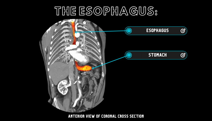
Anatomy of the Esophagus
The esophagus is a muscular tube that carries liquids and food from the pharynx superiorly to the stomach inferiorly. As the esophagus travels inferiorly within the thoracic cavity it sits slightly left of center until the 5th thoracic vertebra. Here the esophagus becomes centrally located and, at the level of the 10th thoracic vertebra, the esophagus shifts anteriorly and slightly left and enters the esophageal opening of the central tendon.
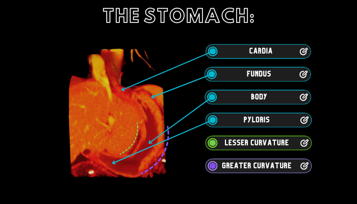
External Anatomy of the Stomach
Because one or more segments of the stomach may be involved in a hiatal hernia, it is important to understand the various segments of the stomach. Externally, the stomach possesses the following segments:
-
The esophagus joins with the stomach at the cardia of the stomach.
-
The fundus is the segment of the stomach that extends superiorly to the cardia.
-
The body of the stomach is the largest segment of the stomach and possesses two curvatures: the greater curvature and the lesser curvature.
-
The inferior, narrowing segment of the stomach is termed the pylorus. The pylorus is subdivided into the pyloric antrum and the pyloric canal.
Anatomy of a Hiatal Hernia
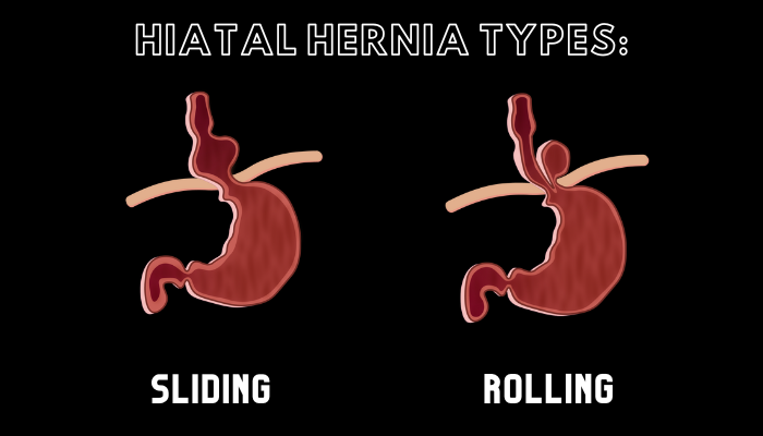
Although a hiatal hernia may be congenital, most occur later in life, as the esophageal opening of the central tendon of the diaphragm weakens and widens. Two types of hiatal hernias are described in the medical literature: sliding hernia and rolling hernia (also termed a paraesophageal hernia).
With a sliding hernia, which is the most common form of hiatal hernia, an upper segment of the stomach and a lower segment of the esophagus slide superiorly into the thoracic cavity through the esophageal opening of the central tendon when the patient lies down or bends over. When the patient regains the upright position both segments slide inferiorly back into the abdominal cavity.
In the rolling hernia, the junction between the esophagus and the cardia of the stomach remains in their normal anatomical positions. However, the fundus of the stomach rolls superiorly through the esophageal opening of the central tendon into the thoracic cavity, coming to lie anteriorly to the lower thoracic segment of the esophagus.
Symptoms of a Hiatal Hernia
Most individuals that have a hiatal hernia do not feel the hernia itself. In addition, these individuals typically do not notice an external bulge in either the thoracic or abdominal regions. Symptoms of a hiatal hernia are mostly related to chronic acid reflux, also termed gastroesophageal reflux disease, or GERD. GERD results from the abnormal position of the stomach and esophagus in a hiatal hernia. This causes the passage of gastric juice superiorly into the esophagus. Symptoms of GERD include but are not limited to
-
Difficulty in swallowing
-
Burping and regurgitation of food into the throat
-
Sore throat or hoarseness
-
Heartburn, especially if one lies down after a meal
-
Chest pain not related to any cardiac pathologies
-
Indigestion
-
Nausea
-
Shortness of breath
-
Pain in the lower chest or upper abdomen, especially after meals
Complications From and Diagnosis of a Hiatal Hernia
Although most hiatal hernias are not severe, they should be regularly monitored by a physician to prevent serious complications and damage to the esophagus and/or stomach. Complications of a hiatal hernia may include
-
Tissue necrosis is due to compression of blood vessels supplying the esophagus or stomach. Although rare, this is a serious complication of a hiatal hernia that typically requires surgery.
-
Esophagitis is inflammation of the esophagus due to irritation from GERD.
-
Constriction of the esophagus due to inflammation of the esophagus and the development of associated scar tissue.
-
Development of esophageal cancer due to chronic irritation from gastric acids entering the esophagus.
-
Gastritis is inflammation of the stomach due to trapped gastric acid within one or more segments of the stomach.
Diagnosis of a hiatal hernia is typically accomplished by a complete physical and a variety of testing procedures, including x-rays and an upper endoscopic exam of the esophagus.
Treatment of a Hiatal Hernia
The most common treatment for a minor hiatal hernia is termed “wait and watch”. Here the patient will undergo frequent tests to monitor whether or not the hernia has worsened. If the hernia progresses your physician might prescribe one of several types of medications to lessen the effects and symptoms of the GERD that frequently accompanies a hiatal hernia.
Surgical repair of a hiatal hernia is rare. However, it will be recommended if the patient’s quality of life is significantly impaired if the hernia symptoms do not respond to any form of treatment, or if the nature of the hernia indicates serious and permanent damage to the esophagus, stomach, and/or diaphragm. Surgical repair varies from noninvasive laparoscopic repair to invasive open surgical repair. The goal of any form of surgical repair is to repair the esophageal opening in the diaphragm and to ensure that the esophagus and stomach remain in their normal anatomical positions, thereby preventing permanent damage to any of the structures involved in the hiatal hernia.
Hiatal hernias typically do not cause noticeable symptoms. However, if they do most individuals are able to alleviate the symptoms with medication and or lifestyle changes. Surgical repair for such a condition is rare but, if needed, often eliminates all signs and symptoms, and prevents a reoccurrence of the hiatal hernia.
Key Terms
GERD - Also termed gastroesophageal reflux disease, GERD results from the abnormal position of the stomach and esophagus in a hiatal hernia. This abnormal position causes the passage of gastric juice superiorly into the esophagus.
Gastritis - Inflammation of the stomach due to trapped gastric acid within one or more segments of the stomach.
Umbilical hernia - A type of hernia where fat or a portion of either the small or large intestine enters into the inguinal canal of the pelvic wall.
Cardia of the stomach - The segment of the stomach where the stomach and esophagus meet.
Rolling hernia - A type of hiatal hernia where the junction between the esophagus and the cardia of the stomach remain in their normal anatomical positions. However, the fundus of the stomach rolls superiorly through the esophageal opening of the central tendon into the thoracic cavity, coming to lie anteriorly to the lower thoracic segment of the esophagus.
Esophageal opening of the diaphragm - One of the three openings in the diaphragm. This opening is located in the anterior, left segment of the central tendon. In addition to the esophagus both vagus nerves and branches of the left gastric artery and vein pass through this opening.
Sliding hernia - The most common form of hiatal hernia. Here an upper segment of the stomach and a lower segment of the esophagus slide superiorly into the thoracic cavity through the esophageal opening of the central tendon when the patient lies down or bends over. When the patient regains the upright position both segments slide inferiorly back into the abdominal cavity.
Esophagitis - Inflammation of the esophagus due to irritation from GERD.
Vertebral portion of the diaphragm - This portion of the muscular segment of the diaphragm has its origin on the anterior surfaces of the first three lumbar vertebrae and their associated intervertebral discs.
Schedule a demo today to learn how you can incorporate BodyViz into your classes and give your students the opportunity to practice using the information from this Brain Builder on real patients with authentic 3D dissection!
Schedule a Demo
Helpful Links: