Tissue Types
by Susan Terry, MS | July 30, 2024

Video explaining the tissue types of the body with a patient example that showcases the skin reparation process!
Use the button below to schedule a demo to learn about our activities, flash cards, and other anatomy resources that support this Brain Builder!
Schedule a Demo
Overview
We often do not think about the tissues of our body until we read an article or see a video about someone who had a terrible accident or someone who has a rare disorder that affects their skin, muscles, or nerves. The human body’s cells are specialized into different tissues. Each of the four types of tissue performs different functions. When cells wear out or you get injured, the cells divide and replace themselves in most tissues.
Background Information
The human body is composed of four different types of tissues. A tissue is a group of cells that work together to perform a common function. The four types of tissues are epithelial tissues, connective tissues, muscle tissues, and nervous tissues. In this article, we will take an in-depth look at epithelial and connective tissues and give an overview of muscle and nervous tissues to tie it all together with information on how the skin repairs itself from wounds.
Core Concepts
Epithelial tissues are classified by the number of cell layers present and the shape of the cells in the tissue. Simple epithelia have only one layer of cells. The cell types are squamous, cuboidal, and columnar. Stratified epithelia have more than one layer of cells and are found in areas that experience a lot of friction, such as the epidermis and the lining of the mouth and throat.
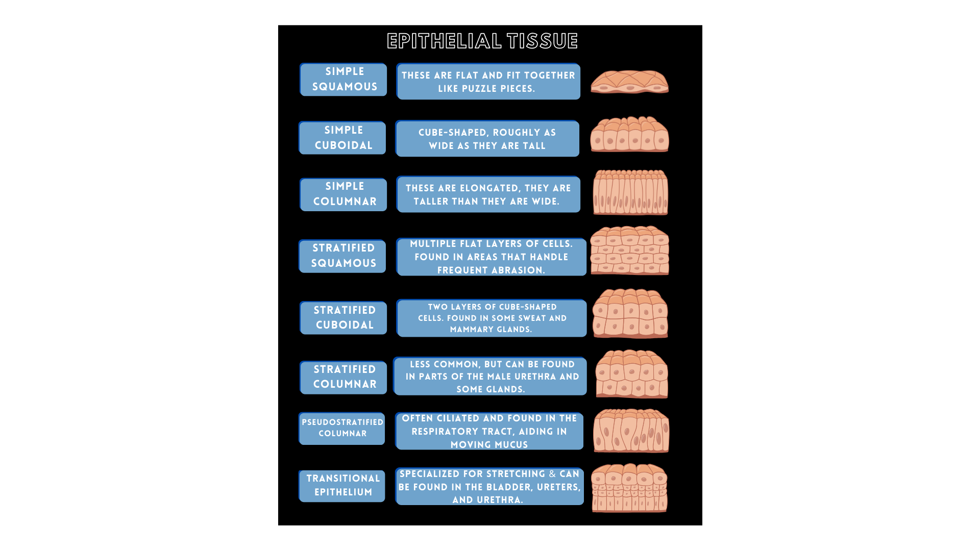
Notice the squamous cells are flat and fit together like puzzle pieces. Cuboidal cells look like small cubes and are about as wide as tall. Columnar cells are elongated and taller than they are wide. The epithelial tissue is connected to the connective tissue underneath it by a basement membrane.
Epithelial tissues serve multiple vital functions within the body, including covering and protecting structures, absorbing nutrients, secreting substances, filtering, excreting, facilitating diffusion, and aiding in touch reception.
- Cover and protect - the skin is made of epithelial tissue that covers and protects the underlying organs from damage and from drying out.
- Secretion - sweat glands are made of epithelial tissue and they secrete sweat
- Filtering - the epithelial tissue of the kidneys filters nitrogenous wastes out of the blood
- Excretion - epithelial cells that have goblet cells produce mucus and release it onto their surface
- Diffusion - the simple squamous epithelium in the alveoli of the lungs allows oxygen to diffuse into the blood and carbon dioxide to diffuse out of the blood
- Touch reception - the epithelial tissue of the skin has sensory receptors in it that help sense touch and pressure
When injured, epithelial cells act swiftly to repair damaged areas, aiding in wound healing and tissue restoration. Study the table below to see where the different types of epithelial tissues are found and what functions they perform.
Connective Tissue
Connective tissues vary widely in composition and are widespread in the body. The functions of connective tissues include supporting and cushioning organs and cells, transporting blood gasses, transporting nutrients, defending against invading organisms, and helping repair damaged tissues. Connective tissues have a nonliving matrix, which is also known as a ground substance. The matrix is an inorganic portion in which cells are embedded and Its consistency can range from liquid to stony hard. They also have protein fibers present. Connective tissues vary in the quantity and quality of their blood supply, which affects how well they regenerate and heal. Some types of connective tissue have a good blood supply such as bone, while ligaments and tendons have a limited blood supply and cartilage has no blood supply at all.
There are two main connective tissue types: proper and specialized connective tissue. Let’s look at the different types of connective tissue in more detail.
Connective tissue proper comprises two main types: loose connective tissue and dense connective tissue. (The only type of loose connective tissue is areolar tissue.) This tissue binds organs together, offering flexibility due to the considerable space between its protein fibers
Dense regular connective tissue includes tendons, ligaments, and aponeuroses (which are connective tissue sheets such as the one that covers the top of the skull). These tissues contain densely packed protein fibers that connect and stabilize structures. Collagen and elastin are two types of fibers found in dense regular connective tissue.
Dense irregular connective tissue, as seen in the dermis, does not have protein fibers that are well arranged. This type of tissue protects underlying organs from damage and resists ripping and tearing. This type of tissue is also seen in the sclera of the eye, the periosteum (which covers long bones), and the outer lining of the testis.
Specialized connective tissue includes any type of connective tissue with specialized cells or specialized ground substance. Examples include:
- adipose tissue: fat droplet
- bone tissue, which has a hard stony matrix with cells embedded in cavities
- blood, which has living cells in its nonliving matrix called plasma.
Muscular Tissue
Muscles are important to the body because they create movement in conjunction with the skeletal system. Muscles also give support to the body, circulate blood, and generate heat. The functions of muscular tissue encompass excitability, extensibility, contractility, and elasticity.
- Excitability means that they can respond to stimuli from nerve impulses or hormones.
- Extensibility means that they can be stretched and extended.
- Elasticity means that they return to their original length when the force is released.
- Contractility means muscle tissue can pull and forcibly shorten.
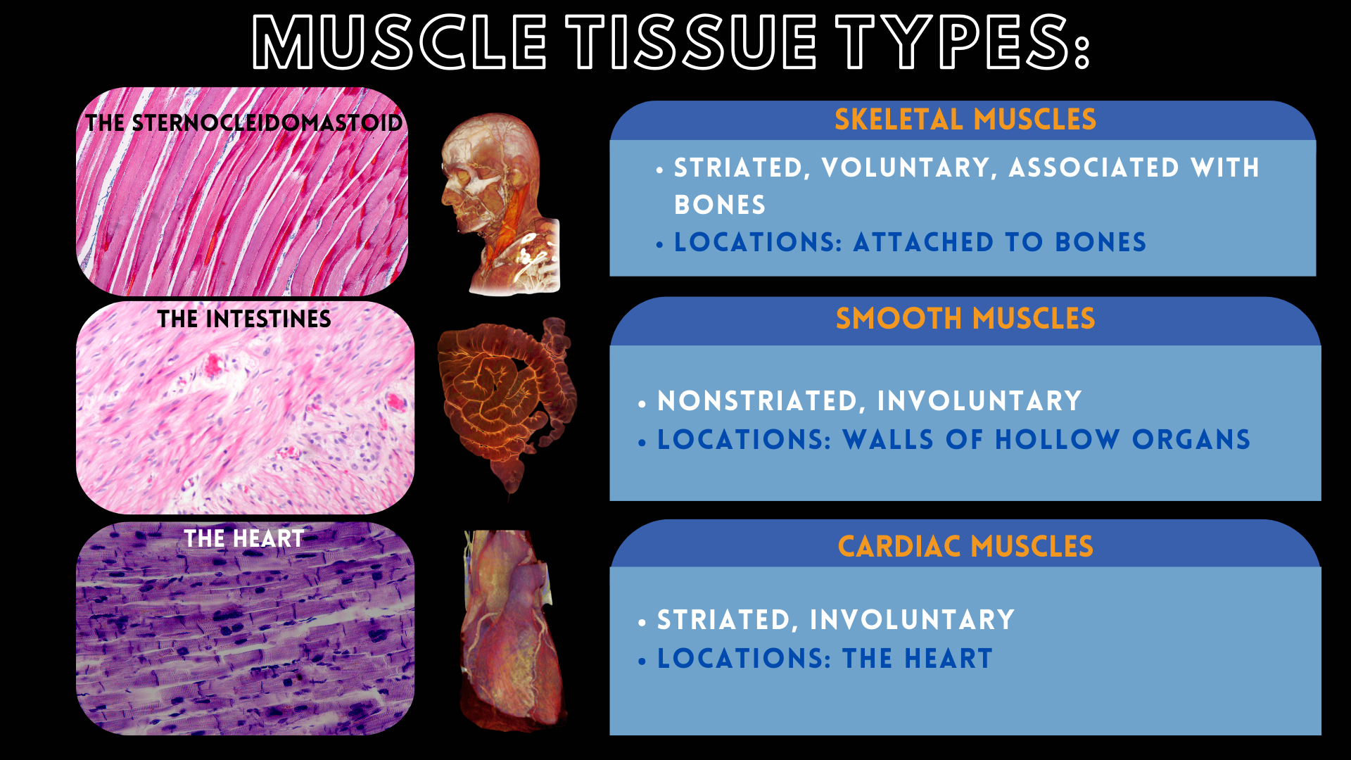
Muscle tissue is made of actin and myosin fibers. The three types of muscle tissue are; skeletal muscle tissue, smooth muscle tissue, and cardiac muscle tissue. Skeletal muscle tissue is characterized by its striated appearance, resulting from the alternating arrangement of actin and myosin fibers. Skeletal muscle tissue is voluntary, so it depends on a signal from the nervous system to work. Smooth muscle is considered to be nonstriated, so the actin and myosin fibers are not arranged in alternating bands and smooth muscle is involuntary. Smooth muscle is found as a part of hollow organs. Cardiac muscle is found only in the heart. It is striated and is involuntary. Its whole purpose is to pump blood throughout the body.
Nervous Tissue
Nervous tissue is composed of neurons and neuroglia and makes up the brain, spinal cord, and all the nerves in the body. Neurons consist of a cell body with dendrites, which are cytoplasmic extensions that receive stimuli from other neurons. The end opposite the dendrites has a long extension called an axon. At the end of the axon are small extensions called axon terminals or synaptic buttons, where a neurotransmitter is released. The neurotransmitter diffuses across the synaptic cleft and attaches to the dendrites of the next axon.
Neurons are responsible for sending electrical signals that coordinate and control the body and its parts. A few examples of nervous tissue functions include muscle contractions, regulation of digestion, and responses to stimuli.
Repairing Damaged Tissues
How well tissues repair themselves is heavily influenced by their blood supply. Nervous tissue does not regenerate. Consequently, a nervous tissue injury could mean long-term incapacitation. Cartilage, being avascular, and ligaments and tendons, which have limited blood supply, face challenges in the healing process. If you know anyone who has injured their cartilage or tore a ligament or tendon, they likely required surgery or experienced prolonged recuperation periods.
Most muscle tissues do not regenerate well. While smooth muscle can regenerate, skeletal and cardiac do not possess significant regenerative abilities. People who suffer myocardial infarction have scar tissue that forms in their heart muscle. Surgery that involves cutting through muscle often involves scar tissue, although a good surgeon will do everything possible to reduce the impact of scarring.
Now, let’s take a look at the regeneration process of epithelial tissues.
- Hemostasis: After a wound occurs, chemicals in the blood signal platelets to start plugging the wound and stopping the blood flow.
- Inflammation: White blood cells arrive to take care of any debris or bacteria.
- Proliferation: Small blood vessels and new skin begins to form. This tissue is pink and delicate.
- Remodeling: Over weeks or months, the skin becomes stronger, and collagen fibers are added to the tissue which increases the strength. Scar tissue may form if the wound is large, jagged, or very deep.
Most small scratches and cuts can be handled at home with washing, some antibiotic ointment, and a small bandage. But what happens if your cut is very deep or very large? You might need some help from a medical professional.
When you appear at the doctor’s office or emergency room with a large cut, the doctor will first make sure the bleeding is under control. They will inspect the cut and thoroughly clean it. Then, the doctor must decide what method they will use to close the cut, so it will heal – hopefully with as minor a scar as possible. If it is very deep and depends on the part of the body affected, the doctor may send you for imaging studies to make sure no underlying organs are affected and that there are no broken bones.
The closure options are sutures, staples, adhesive strips, and surgical glue. A small straight cut might be closed with surgical glue. This is often used in arthroscopic surgeries. A deep but straight cut might get staples or be secured with adhesive strips. Orthopedic surgeries involving longer incisions may require staples or sutures.
Key Terms:
Tissue – a group of cells that work together to perform a common function
Simple epithelium – epithelial sheet with one layer of cells
Stratified epithelium – epithelial sheet with more than one layer of cells
Basement membrane – a thin layer of protein fibers present under a layer of epithelium
Squamous – refers to cells that are flat and fit together like puzzle pieces
Cuboidal – refers to cells that look like cubes with a nucleus in the center
Columnar – refers to cells that are taller than they are wide with a nucleus in the middle.
Pseudostratified – refers to a type of epithelial sheet that looks like it may be stratified because of the irregularity of its cell shapes, but is only one layer
Connective tissue – body tissue with cells, ground substance, and fibers that perform a wide range of functions
Matrix – the inorganic portion of connective tissue where cells are embedded which can be liquid, gel-like or stony hard
Ground substance – gel like inorganic matrix material
Collagen – protein fibers in tissues that allow stretching without injury
Elastin – protein fibers in tissues that allow the tissue to spring back to its original position when stretched
Reticular fibers -protein fibers that form a mesh-like network and are seen in lymph nodes
Schedule a demo today to learn how you can incorporate BodyViz into your classes!
Schedule a Demo
Helpful Links: